 Jul, 3 2026

You twist your hips during a soccer drill or pivot hard on the basketball court, and suddenly, there it is-a sharp catch in the front of your groin. It’s not a muscle strain you can shake off with ice and rest. For athletes, this specific type of pain often points to a hip labral tear, which is damage to the fibrocartilaginous rim that lines the acetabular socket of the hip joint. This small but critical structure acts as a seal for the hip joint, keeping everything stable and lubricated. When it tears, the consequences can sideline even the most dedicated competitor.
If you are an athlete dealing with persistent hip pain, understanding the path from injury to recovery is crucial. You need to know why standard MRIs might miss the problem, when surgery becomes necessary, and what realistic timelines look like for getting back into the game. This guide breaks down the medical reality of hip labral tears, focusing on the latest diagnostic standards and surgical outcomes for active individuals.
Understanding the Anatomy and Causes
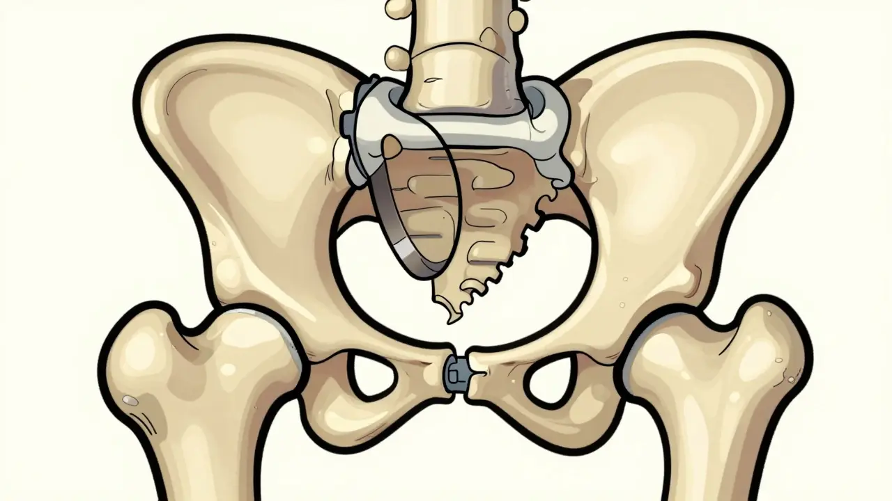
To fix the hip, you first have to understand what broke. The hip joint is a ball-and-socket design. The "ball" is the head of the femur (thigh bone), and the "socket" is the acetabulum in the pelvis. Lining the edge of that socket is the labrum, which is a ring of cartilage that deepens the socket and provides suction stability. Think of it like the gasket on a jar lid; if the gasket is torn, the seal is broken, and pressure escapes.
Most athletic labral tears don’t happen from a single traumatic event like a car crash. Instead, they are usually caused by femoroacetabular impingement (FAI), which is a condition where abnormal bone growth causes the femoral head and acetabulum to rub against each other abnormally. Imagine two gears that aren’t perfectly aligned grinding together every time you move. Over thousands of repetitions in sports like hockey, gymnastics, or running, this friction pinches and eventually tears the labrum.
This issue is incredibly common in young, competitive athletes. Research indicates that between 22% and 55% of athletic hip pain cases involve labral pathology. If you are under 40 and play a sport requiring repetitive hip rotation, you are in the highest-risk demographic. Ignoring the pain doesn’t make it go away; untreated labral tears are associated with a 4.5 times higher risk of developing osteoarthritis within ten years.
The Diagnostic Challenge: Why Standard MRIs Fail
One of the biggest frustrations for athletes is walking out of a doctor’s office with a normal MRI result despite having severe pain. Here is the catch: conventional MRI scans have only 35% to 60% sensitivity for detecting labral tears. They simply aren’t designed to see soft tissue details inside the joint capsule clearly enough.
The gold standard for non-invasive diagnosis is magnetic resonance arthrography (MRA), which is an MRI procedure where contrast dye is injected directly into the hip joint to highlight tears. By filling the joint with dye, radiologists can see exactly where the labrum separates from the bone. MRA boasts a 90% to 95% sensitivity rate. Recent advancements in 2023 now include 3D MRI sequencing, which pushes diagnostic accuracy up to 97% for complex cases.
Before any advanced imaging, doctors will start with plain X-rays. These are essential not to see the tear itself, but to check the bone structure. They look for dysplasia (shallow sockets) or cam/pincer deformities (bone spurs). Physical exam tests like FADIR (flexion, adduction, internal rotation) also play a huge role. If bending your knee toward your chest while twisting inward reproduces your pain, you have a strong clinical indicator of a labral issue.
| Method | Sensitivity/Accuracy | Primary Purpose | Limitations |
|---|---|---|---|
| Plain Radiographs (X-Ray) | N/A (Structural only) | Check for bone abnormalities (FAI, Dysplasia) | Cannot visualize soft tissue or cartilage tears |
| Conventional MRI | 35% - 60% | Rule out other conditions (fractures, tumors) | Misses up to 65% of labral tears |
| Magnetic Resonance Arthrography (MRA) | 90% - 95% | Visualize labral detachment and cartilage damage | Invasive (requires injection); higher cost ($1,200-$1,800) |
| Hip Arthroscopy | 98% | Definitive diagnosis and simultaneous treatment | Surgical risks; requires anesthesia and recovery time |
Conservative Treatment: Can You Avoid Surgery?
Not every labral tear requires an operation. Many athletes try conservative management first, especially if the tear is partial or if symptoms are manageable. The typical protocol involves 4 to 6 weeks of relative rest, avoiding movements that cause pinching, along with NSAIDs like ibuprofen to reduce inflammation.
Physical therapy is controversial but often effective. While some older studies suggested only 30% success rates with PT alone, newer data from specialized sports clinics shows that about 65% of athletes can manage their symptoms without surgery through targeted strengthening. The goal isn’t just to rest the hip; it’s to strengthen the glutes and core to stabilize the pelvis and reduce stress on the joint.
Corticosteroid injections can provide temporary relief for 3 to 6 months in 70% to 80% of cases. However, these are generally used as a diagnostic tool or a bridge to surgery, not a long-term cure. Recently, regenerative medicine has entered the scene. Platelet-rich plasma (PRP) injections showed promise in a 2022 randomized controlled trial, with 55% of patients avoiding surgery at the 12-month mark. While not covered by all insurance plans, PRP offers a middle ground between doing nothing and cutting into the joint.
If you haven’t seen significant improvement after three to six months of conservative care, or if mechanical symptoms (catching, locking) persist, surgery becomes the standard recommendation.
Hip Arthroscopy: The Surgical Solution
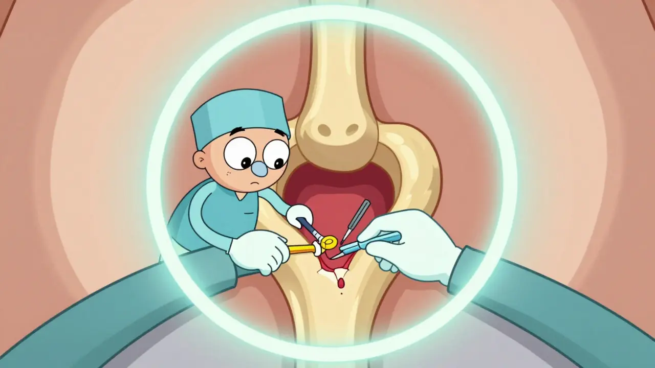
When non-surgical options fail, hip arthroscopy, which is a minimally invasive surgical procedure using a camera and small instruments to repair the hip joint, is the next step. This isn’t open surgery with large incisions. Surgeons make tiny portals around the hip, insert a camera, and work inside the joint.
There are two main types of procedures performed during arthroscopy:
- Debridement: The surgeon trims away the torn, unstable fragments of the labrum. This is quicker and has a shorter recovery time (3-4 months), but it removes tissue that helps seal the joint. It’s often reserved for degenerative tears or older patients who won’t benefit from repair.
- Labral Repair: The surgeon reattaches the labrum to the acetabular rim using suture anchors. This preserves the joint’s natural mechanics. Recovery takes longer (5-6 months), but it offers better long-term protection against arthritis.
A critical factor in surgical success is addressing the underlying cause. If you have FAI, the surgeon must shave down the excess bone (osteoplasty) along with repairing the labrum. Leaving the bone spur in place guarantees the new repair will fail. Similarly, if you have hip dysplasia, isolated labral repair has a 65% failure rate. In those cases, a more extensive pelvic osteotomy might be required alongside or instead of arthroscopy.
Technology is advancing rapidly here too. In June 2023, the FDA approved the first bioabsorbable suture anchor system specifically for labral repair. Early data shows an 89% success rate at two years, compared to 82% for traditional metal or plastic anchors. These dissolving anchors eliminate the need for future removal surgeries and may reduce irritation.
Recovery and Return to Sport
Getting off the operating table is only half the battle. Rehabilitation for hip arthroscopy is rigorous and follows a strict timeline. Most protocols span six months and are divided into phases:
- Weeks 1-6 (Protection): You’ll likely use crutches. The focus is on reducing swelling and restoring basic range of motion without stressing the repair. Weight-bearing is limited.
- Weeks 7-12 (Strengthening): Crutches come off. You begin progressive strengthening of the hip abductors, extensors, and core. Gait training ensures you walk normally again.
- Weeks 13-20 (Sport-Specific Training): Agility drills, plyometrics, and sport-specific movements are introduced. This phase builds endurance and neuromuscular control.
- Weeks 21-26 (Return to Sport): Full participation resumes once criteria are met.
What are those criteria? Experts agree you shouldn’t return until you have achieved 90% quadriceps strength symmetry compared to your uninjured leg and can perform pain-free hip internal rotation to 30 degrees. Professional athletes like NHL player Ryan Nugent-Hopkins took about 5.5 months to return to professional hockey after a labral repair.
Outcomes are generally positive. Competitive athletes demonstrate an 85% to 90% return to pre-injury sport levels following arthroscopic intervention. However, this drops to 70% to 75% for athletes over 35 years old. Sports requiring extreme ranges of motion, such as ballet or gymnastics, carry a 25% higher complication rate post-surgery due to the high demands placed on the repaired tissue.
Risks, Costs, and Realistic Expectations
No surgery is without risk. Common complications include persistent pain (15-20% of cases), heterotopic ossification (abnormal bone growth, 5-10%), and nerve injury (1-2%). Revision surgery rates sit at 8-12% at five years, often due to incomplete initial diagnosis or untreated structural issues.
Cost is another major barrier. While standard MRIs might cost $500-$800, MRAs often run $1,200-$1,800 and are frequently paid out-of-pocket. Insurance approvals for hip arthroscopy can also be difficult, requiring extensive documentation of failed conservative treatments. Athletes with access to specialized sports medicine centers report 92% satisfaction rates with outcomes, compared to 75% at general orthopedic practices, highlighting the importance of choosing a surgeon who specializes in hip preservation.
How long does it take to recover from hip labral tear surgery?
Recovery typically takes 5 to 6 months for a full return to sport after a labral repair. Debridement procedures may allow a return in 3 to 4 months. The rehabilitation process involves protected weight-bearing for the first 6 weeks, followed by progressive strengthening and sport-specific training.
Can hip labral tears heal without surgery?
Yes, approximately 65% of athletes can manage symptoms through conservative treatment including physical therapy, activity modification, and possibly PRP injections. However, if mechanical symptoms like catching or locking persist, or if pain continues after 3-6 months of therapy, surgery is usually recommended to prevent further joint damage.
What is the best imaging test for a hip labral tear?
Magnetic Resonance Arthrography (MRA) is considered the gold standard for non-invasive diagnosis, offering 90-95% sensitivity. Standard MRIs often miss these tears, showing only 35-60% sensitivity. Plain X-rays are also essential to check for underlying bone abnormalities like FAI or dysplasia.
Does hip arthroscopy prevent osteoarthritis?
Hip arthroscopy aims to preserve joint function and delay the onset of osteoarthritis. Untreated labral tears are associated with a 4.5 times higher risk of developing hip OA within 10 years. While surgery repairs the current damage, it does not guarantee complete prevention of arthritis, especially if underlying structural issues like dysplasia are not addressed.
What sports are hardest to return to after hip labral repair?
Sports requiring extreme hip rotation and flexion, such as ballet, gymnastics, and hockey, present the greatest challenges. These activities have a 25% higher complication rate post-surgery due to the high mechanical demands placed on the repaired labrum. Athletes in these sports require meticulous rehabilitation and careful monitoring before returning.